(7 of 38)
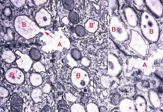
TEM of Serous Acinus
This electron micrographs shows the lumen of a serous acinus (A) in the parotid gland of a mouse. Note the secretory granules (B) that fill the apical region of each serous cell bordering the lumen. In the image on the left, one of these granules is being extruded into the lumen (arrow) in a merocrine fashion. All salivary glands are classified as merocrine glands.

Legend
|
A - lumen of serous acinus |
B - serous secretory granules |