(5 of 38)
Serous Secretory Acini
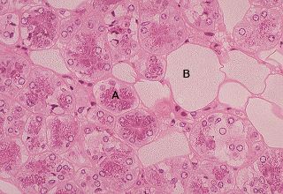
This image is a high power photomicrograph taken of a plastic-embedded piece of the parotid gland. The field is filled with serous secretory acini (A) and fat cells (B). Plastic embedding facilitates resolution of cellular detail. It is evident that the apical region of each serous acinar cell is filled with acidophilic secretory granules of varying size. Note the shape and size of the nucleus in each cell.

Legend
|
A - serous secretory acinus |
B - fat cell |