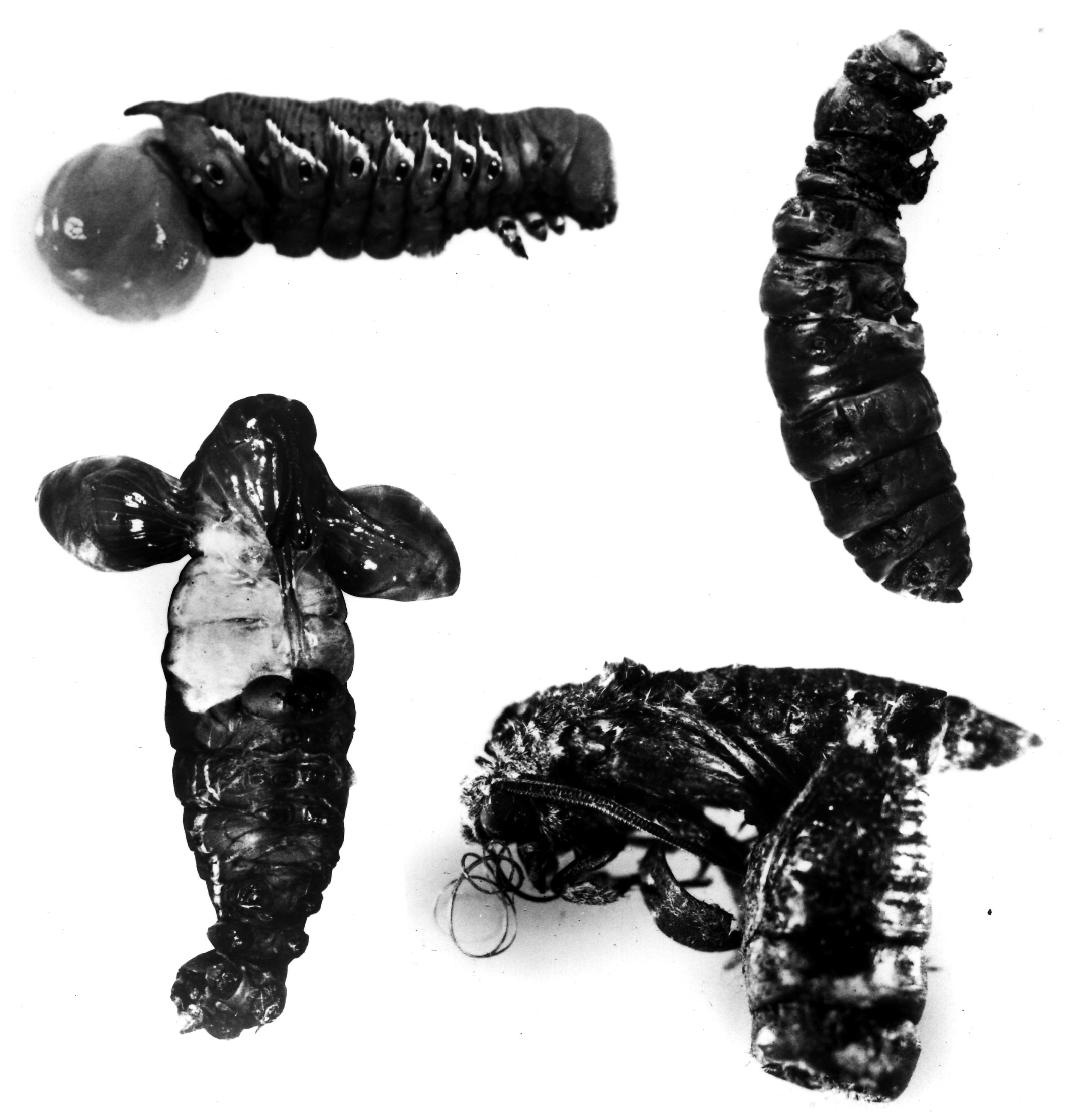
As documented previously, the higher plant nonprotein amino acid L-canavanine is a potent arginine antimetabolite that is typically insecticidal to non-adapted insects (Rosenthal, 1991). For example, terminal instar larvae of the tobacco hornworm, Manduca sexta [Sphingidae], reared on an agar-based diet supplemented with 2.5 mM canavanine, produced pupae and adults that exhibited striking developmental aberrations.

BIOLOGICAL CONSEQUENCES of canavanine consumption by the tobacco hornworm, Manduca sexta. Top left, larvae given 1.0 mg/g fresh body weight L-canavanine exhibit severe diuresis and often the gut is everted through the anus; top right and bottom left, pupal developmental aberrations resulting from rearing terminal instar larvae on 2.5 mM canavanine-containing diet; bottom right, adult malformations resultign from the rearing conditions described previously. (Photo provided by D.L. Dahlman).
In marked contrast, larvae of the tobacco budworm, Heliothis virescens [Noctuidae], a highly destructive agricultural pest (Lincoln, 1972) reared on canavanine-containing diet had an LC50 value for this arginine antagonist of 300 mM (Berge et al., 1986).This LC50 corresponds to 53,000 ppm wet diet weight or nearly 40% on a dry weight basis (Berge et al., 1986). While the pupae that emerged from these canavanine-treated larvae were depauperate, they lacked discernible developmental aberrations. A group of five larvae sustained on a staggering 500 mM canavanine-containing diet survived for 9 days before the first larval dead was recorded.

Heliothis virescens resistance to this arginine antagonist was also demonstrated by parenteral injection of canavanine: the LD50 for this nonprotein amino acid in M. sexta was 1.0 g kg-1 fresh body weight as compared to 10.7 g kg-1 for H. virescens (Berge et al., 1986). Heliothis virescens might tolerate elevated levels of dietary canavanine because of its ability to excrete this potentially toxic allelochemical. Analysis of the larval fecal matter, produced by terminal instar larvae from feeding on 150 mM canavanine-containing diet, disclosed that only 0.6% of the consumed canavanine was eliminated by this excretory route. Examination of the body fluids and tissues established the lack of significant sequestration of canavanine within the larval body (Berge and Rosenthal, 1990). Thus, H. virescens actively metabolized canavanine; the t1/2 for canavanine clearance from the hemolymph was 135 min (Berge and Rosenthal, 1990).
LARVAL ABILITY TO TOLERATE CANAVANINE
Biochemical studies were conducted to determine if larval ability to tolerate canavanine resulted from its constitutive metabolism, or alternatively was induced in response to dietary consumption of canavanine (Berge and Rosenthal, 1990). In one study, the t1/2 for canavanine clearance of parenterally injected L-[guanidinooxy-14C]canavanine, in larvae maintained on a 150 mM canavanine-containing diet for 72 h, was 138 min. This is exactly the same value found for control insects that were not exposed previously to canavanine prior to determining the t1/2 value for canavanine clearance.
In another approach to resolving this question, H. virescens larvae were provided sufficient cycloheximide to inhibit nearly 80% of the L-[3H]leucine-labeled protein formation observed in control animals. Such cycloheximide-treated insects cleared canavanine with a t1/2 equal to 121 min; once again this value was identical in control animals (Berge and Rosenthal, 1990). Electrophoretic analysis of hemolymph proteins of larvae reared on 150 mM canavanine-containing diet was identical to that obtained from larvae raised on control diet.
IN SUMMARY, NONE OF THE DESCRIBED EXPERIMENTS PROVIDED EVIDENCE FOR SYNTHESIS OF A UNIQUE PROTEIN BY CANAVANINE-TREATED INSECTS. THIS RESULT SUPPORTED THE VIEW THAT H. VIRESCENS DREW UPON A PREEXISTING, CONSTITUTIVE ENZYME TO CATABOLIZE CANAVANINE.
Analysis of the various body organs of the larvae demonstrated that canavanine catabolism occurred solely in the gut. In particular, the fat body, the rough insectan equivalent of the mammalian liver and a major detoxification organ, did not possess significant canavanine-degrading ability. Moreover, the canavanine-catabolizing enzyme of the larvae was found exclusively in the gut, and was probably part of the gut wall rather than secreted into the gut lumen since this enzyme was not lost when the gut contents were removed by thorough washing. the microbody fraction of an extract of the larval gut could not degrade canavanine; this ability resided solely in the soluble fraction of the gut extract.
CANAVANINE CATABOLISM
To unravel the metabolic disposition of canavanine, the larvae were
provided L-[guanidinooxy-14C]canavanine. The principal
degradation product of radiolabeled canavanine was [14C]guanidine(Berge
and Rosenthal, 1991). Subsequent in vivo studies employing L-[1,2,3,4-14C]canavanine
identified L-[1,2,3,4-14Clhomoserine as the preponderant radiolabeled
catabolite. This finding immediately suggested the following pathway in
which the 14carbon atom of the guanidinooxy moiety of canavanine
was transferred to guanidine:
Other experiments with larvae of H. virescens led to the discovery of a gut enzyme--a larval reductase able to catalyze an NADH-dependent reduction of hydroxyguanidine to guanidine (Rosenthal, 1992). This finding created the intriguing possibility that canavanine was catabolized initially to homoserine and hydroxyguanidine rather than guanidine by a novel hydrolase able to cleave the O-N bond of the guanidinooxy moiety of the substrate.

While this metabolic ability has been observed in a soil-borne Pseudomonas (Kalyankar et al., 1958), the responsible enzyme was not isolated nor has this metabolic capacity been described from an eukaryotic organism.
THE EXPERIMENTAL EVIDENCE INDICATED TWO ENZYMES WERE FUNCTIONING IN CONCERT TO METABOLIZE CANAVANINE.
![]() THE
FIRST REACTION EMPLOYED A NOVEL HYDROLASE THAT DIRECTED THE FORMATION OF
HOMOSERINE AND HYDROXYGUANIDINE FROM CANAVANINE.
THE
FIRST REACTION EMPLOYED A NOVEL HYDROLASE THAT DIRECTED THE FORMATION OF
HOMOSERINE AND HYDROXYGUANIDINE FROM CANAVANINE.
![]() THE
SECOND REACTION, HYDROXYGUANIDINE WAS REDUCED TO GUANIDINE.
THE
SECOND REACTION, HYDROXYGUANIDINE WAS REDUCED TO GUANIDINE.
CANAVANINE HYDROLASE
The existence of canavanine hydrolase (CH), an enzyme able to cleave an oxygen-nitrogen bond, would be an important finding since this enzyme is the only protein known to demonstrate this ability. As such it would represents a new type of hydrolase--one that acts on oxygen-nitrogen bonds (EC 3.13.1.1).
The search for this enzyme in the gut of larval H. virescens culminated in the isolation of a homogeneous enzyme that mediated an irreversible hydrolysis of L-canavanine to L-homoserine and hydroxyguanidine (Melangeli et al., 1997). Canavanine hydrolase (EC 3.13.1.1) exhibited a high affinity for canavanine as judged by the apparent Km value of 1.1 mM for canavanine. The turnover number for this reaction was 21.1 mmol min-1 mmol-1. Canavanine hydrolase also exhibited a high degree of specificity for L-canavanine as it could not function effectively with either L-2-amino-5-(guanidinooxy)pentanoate or 2-amino-3-(guanidinooxy)propionate, the higher or lower homolog, respectively of L-canavanine nor its methyl ester. Canavanine derivatives such as L-canaline and O-ureido-L-homoserine were not metabolized significantly by canavanine hydrolase.
REFERENCES
Berge, M. A., Rosenthal, G. A. and Dahiman, D. L. (1986) Pestic. Biochem. Physiol. 25, 319-326.
Berge, M.A. and Rosenthal, G.A. (1990) J. Food Agr. Chem. 38, 2061-2065.
Berge, M. A. and Rosenthal, G. A. (1991) Chem. Res. Toxicology 4, 237-240.
Kalyankar, G D., Ikawa, M. & Snell, A. A. (1958) J. Biol. Chem. 233, 1175-1178.
Melangeli, C., Rosenthal, G.A. & Dahlman, D.L. (1997) Proc. Natl. Acad. Sci. 94, 2255-2260.
Rosenthal, G. A. (1991) In: Nonprotein Amino Acids as Protective Allelochemicals, eds. Rosenthal, G. A. & Berenbaum, M. (Academic Press, San Diego), pp. 1-11.
Rosenthal, G. A. (1992) Bioorg Chem. 20, 55-61.